
Efficacy of stereomicroscope as an aid in grossing and histopathological diagnosis

*Corresponding author: Rezhat Abbas, Department of Oral Pathology and Microbiology, Government Dental College and Hospital, Srinagar, Jammu and Kashmir, India. writetoempire@gmail.com
-
Received: ,
Accepted: ,
How to cite this article: Abbas R, Dar MS, Latoo SH. Efficacy of stereomicroscope as an aid in grossing and histopathological diagnosis. J Global Oral Health. doi: 10.25259/JGOH_38_2024
Abstract
Objectives
The histopathological analysis of tissues is regarded as the gold standard for identifying lesions. The macroscopic examination of the tissue material is critical to making an accurate histological diagnosis. Grossing is regarded as a necessary but frequently overlooked process in histology. An accurate grossing method reduces errors while also providing useful information about the specimen for a definitive diagnosis. Stereomicroscopic examination of specimens may assist pathologists in providing vital information about the nature of the specimen, which may aid in the correct orientation of the specimen and arriving at the correct diagnosis. With this background, a study was conducted to evaluate the role of stereomicroscope in routine grossing and its contribution to arrive at a definitive diagnosis.
Materials and Methods
The study was conducted on 70 samples of the patients sent to the Department of Oral Pathology, Government Dental College and Hospital, Srinagar. Specimens received were properly labeled and fixed in 10% neutral-buffered formalin for 24 h. All the specimens were observed under the naked eye and stereomicroscope (Radical RXLr-5), where we could appreciate the surface details of the tissue. Based on the stereomicroscopic evidence, they were embedded. The sections were stained with hematoxylin and eosin and observed under a compound microscope.
Results
Compared to the naked eye, the stereomicroscopic observation of specimens showed better details, which helped in the proper orientation of the specimen and aided in the accurate diagnosis.
Conclusion
The stereomicroscope acts as an adjuvant for the proper orientation of the specimen, which helps in arriving at an accurate final diagnosis.
Keywords
Diagnosis
Grossing
Histopathological examination
Oral lesions
Stereomicroscope

INTRODUCTION
The first phase in surgical pathology dissection is “grossing,” which refers to the examination and dissection of surgical specimens, as well as the creation of sections from tissues that need to be processed. An accurate diagnosis is based on the specimen being correctly identified, handled, and processed. Microscopic examination requires proper specimen identification and orientation.[1] An accurate grossing method reduces errors and gives essential information about the specimen for making a reliable diagnosis. However, the importance of this stage is frequently overlooked and ignored. Any error in this critical phase may result in an incorrect diagnosis since incorrect specimen orientation might either cause a delay in diagnosis or create a diagnostic difficulty. If difficulties are encountered when orienting the specimen, photographing the specimen may be beneficial. Imaging methods may include digital photography, stereomicroscopy of the specimen, and even radiographic evaluation.[2] Stereomicroscopic examination of the specimen can aid and provide some essential clues about the form and kind of proliferation (papillary, mucosal, or submucosal, presence of epithelium or capsule), which can help not only with specimen orientation but also with diagnosis.[3] Therefore, we undertook this study to analyze the significance of the stereomicroscope in routine grossing and its contribution to the definitive diagnosis.
MATERIALS AND METHODS
The present prospective study was conducted on 70 samples of patients sent to the Department of Oral Pathology, Government Dental College and Hospital, Srinagar. The received specimens were properly labeled and fixed in 10% neutral buffered formalin for 24 h. The tissues were cleared of blood clots. All the specimens were observed under the naked eye and stereomicroscope [Figure 1] where we appreciated the surface details of the tissue. Based on the stereomicroscopic evidence, they were embedded, and sections were stained with hematoxylin and eosin and observed under a compound microscope. The samples included were soft tissue specimens that include solid tumors, cystic lesions, and surface lesions; both incisional and excisional biopsies; completely fixed tissues in 10% neutral buffered formalin; and tissues with minimal dimensions, fragmented tissues, and tissues with forceps artifacts. The samples excluded were as hard tissue specimens and tissues with incomplete fixation. The interpretation was done as per the criteria laid down by Shashidara et al.,[4] as given in Table 1.
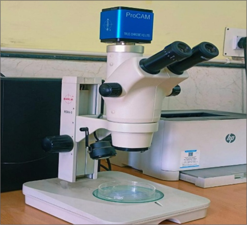
- Stereomicroscope (Radical RXLr-5).
| Colors observed and their interpretations | |
| Color | Interpretation |
| White | Calcification/bone |
| Gray white | Fibrosis |
| Yellow | Lipoma/fat necrosis |
| Brown/black | Hemosiderin/melanin |
| Red | Hemorrhage, blood vessel congestion |
| Consistencies and their interpretations | |
| Consistency | Interpretation |
| Soft | Necrosis, fluid |
| Firm | Fibrosis |
| Hard | Bone, calcification |
| Rubbery | Lymph nodes |
| Cheesy | Keratin, caseous necrosis |
RESULTS
A total of 70 cases were included in the study. There were 40 cystic lesions, 18 solid lesions, and 12 superficial lesions [Table 2]. A significant difference was noted between examination with the unaided eye and stereomicroscope.
| Nature of lesion | Histopathological diagnosis | Number of cases |
|---|---|---|
| Cystic lesions (n=40) | Radicular cyst | 10 |
| Dentigerous cyst | 8 | |
| Odontogenic keratocyst | 6 | |
| Median palatal cyst | 2 | |
| Orthokeratinized odontogenic cyst | 4 | |
| Mucocele | 5 | |
| Buccal bifurcation cyst | 1 | |
| Unicystic ameloblastoma | 2 | |
| Aneurysmal bone cyst | 2 | |
| Total=40 | ||
| Solid lesions (n=18) | Conventional ameloblastoma | 5 |
| Malignant melanoma | 1 | |
| Ossifying fibroma | 3 | |
| Undifferentiated pleomorphic sarcoma | 1 | |
| Squamous cell carcinoma | 6 | |
| Central giant cell granuloma | 2 | |
| Total=18 | ||
| Surface lesions (n=12) | Pyogenic granuloma | 4 |
| Peripheral giant cell granuloma | 2 | |
| Papilloma | 1 | |
| Fibroma | 3 | |
| Peripheral ossifying fibroma | 2 | |
| Total=12 | ||
Grand total=70
The first case was that of a 26-year-old female patient who reported pain and swelling in the lower right back region of the jaw for the past 5 months. Extraorally, the presence of facial asymmetry was noted, with swelling extending anteroposteriorly a few centimeters from the commissure of mouth to the external ear and superoinferiorly from the lower canthus of the eye to the inferior border of the mandible. Intraoral examination showed a bony, hard swelling extending from the lower right canine to the retromolar pad area. Expansion of cortical plates was observed along with the obliteration of the buccal vestibule. The panoramic view revealed multilocular radiolucency on the right side of the mandible, extending from the right side of the ramus of the mandible to the distal surface of the canine. An incisional biopsy was done and the specimen revealed a cystic lesion. Under the stereomicroscope, there was a cystic cavity noted surrounded by a thick wall, and a few intraluminal projections were appreciated. Microscopic examination revealed a cystic cavity lined by odontogenic epithelium with hyperchromatic, palisaded basal layer and stellate reticulum-like suprabasal layers [Figure 2]. A final diagnosis of unicystic ameloblastoma was made.

- (a) Gross picture under naked eye depicting a cystic lesion. (b) Stereomicroscopic image showing cystic lumen (blue arrow) surrounded by fibrous wall (black arrow). (c) Hematoxylin & eosin-stained section (10x) showing fibrous capsule (blue arrow) lined by ameloblastomatous epithelium (black arrow).
The second case was that of a 55-year-old female patient who complained of a growth on the right side of the upper jaw for 6 months. It was gradual in onset, progressive in nature, associated with pain, paresthesia, and bleeding. Intraoral examination revealed an exophytic, pedunculated growth on the right side of the maxillary alveolus extending from the second premolar to the tuberosity area. It was soft in consistency, reddish-blue in color, nodular in shape, and with irregular borders. An excisional biopsy was done, and on gross examination with the naked eye, the specimen was round to oval solid mass. Under the stereomicroscope, it revealed a thin yellowish layer covering a dark brown solid tissue. Microscopic examination showed a parakeratinized stratified squamous epithelium, subepithelial grenz zone, and deeper sections showing numerous multinucleated giant cells in a fibrocellular stroma [Figure 3]. A final diagnosis of peripheral giant cell granuloma was made.

- (a) Gross picture under naked eye, round to oval solid mass. (b) Stereomicroscopic image showing dark brown solid tissue (red arrow) circumscribed by yellowish white layer (black arrow). (c) Hematoxylin & eosin-stained section (10x) showing stratified squamous epithelium (black arrow) and underlying fibrocellular stroma with numerous giant cells (red arrow).
The third case was that of a 32-year-old male patient who reported a swelling in his palate. The swelling was observed four months back. It was insidious in onset and progressive in nature. No pain or discharge was associated with the swelling. Intraoral examination revealed a solitary oval swelling measuring 2 cm × 3 cm in the midline of the palate, 1.5 cm away from the palatal gingival margin. The swelling was soft in consistency and non-tender on palpation. Occlusal radiograph showed symmetrical oval radiolucency in the palate with sclerotic borders. The lesion was behind the incisive canal and corresponded to premolars and molars. Enucleation of the cyst was done under local anesthesia. On gross examination with the naked eye, the specimen was yellowish-white cystic in nature. Under the stereomicroscope, the specimen displayed a cystic cavity surrounded by a fibrous wall. Microscopic examination showed a cystic lumen lined by thin stratified squamous epithelium surrounded by a fibrocellular capsule [Figure 4]. A final diagnosis of median palatal cyst was made.

- (a) Gross picture under naked eye which could not be described well. (b) Stereomicroscopic image showing cystic lumen (red arrow) lined by thin layer of epithelium (black arrow) surrounded by fibrous wall (blue arrow). (c) Hematoxylin & eosin-stained section (10x) showing cystic lumen (red arrow) lined by stratified squamous epithelium (black arrow) and surrounded by fibrocellular capsule (blue arrow).
The fourth case was that of a 76-year-old male patient who reported to the hospital with a complaint of pain, swelling, and bleeding in the upper left posterior tooth region for two months. Intraoral examination revealed a sessile proliferative grayish-brown nodular growth on the left maxillary alveolus with a bosselated surface and multiple bleeding areas measuring approximately 5 cm × 4 cm in size. An incisional biopsy was done and on gross examination, there was a dark brown solid specimen. Under a stereomicroscope, the tissue revealed a degenerated superficial layer and dark brown deeper areas. Microscopic examination revealed numerous atypical melanocytes with features of hyperchromatism, pleomorphism, and altered N/C ratio. Areas of necrosis and degeneration were also noted [Figure 5]. A final diagnosis of malignant melanoma was made.

- (a) Gross picture under naked eye showing solid brown tissue. (b) Stereomicroscopic image showing solid dark brown tissue (red arrow) surrounded by degenerated epithelium (black arrow). (c) Hematoxylin & eosin-stained section (10x) showing atypical melanocytes within a sparse fibrous stroma.
The fifth case is a 15-year-old female patient who reported a swelling on the right side of the lower jaw for three weeks. Intraoral examination revealed a well-defined ovoid swelling present on alveolar mucosa in relation to 45, 46 with normal mucosal color. There was no spontaneous bleeding and no surface ulceration. An excisional biopsy was done, and on gross examination with the naked eye, there was a solid yellowish-white oval mass. Under the stereomicroscope, it revealed a yellowish-white solid mass of tissue with a pale brown covering. Microscopic examination revealed a thin stratified squamous epithelium and underlying dense stroma with spindle-shaped fibroblasts arranged in streaming fascicles [Figure 6]. A final diagnosis of fibromatosis was made.

- (a) Gross picture under naked eye round to oval yellowish white tissue. (b) Stereomicroscopic image showing yellowish white solid tissue (red arrow) surrounded by epithelium (black arrow). (c) Hematoxylin & eosin-stained section (10x) showing stratified squamous epithelium (black arrow) and an underlying stroma showing spindle shaped fibroblasts in streaming fascicles (red arrow).
DISCUSSION
“Grossing” refers to the careful examination and systematic dissection of surgical specimens to produce tissue sections for microscopic examination. It is usually done by a pathologist, resident, physician assistant, histotechnologist, or biomedical scientist. Grossing is the initial step in biopsy reporting that ensures the specimen is handled properly and has been equated with biopsy triage.[4] Histopathological evaluation of tissues is regarded as the gold standard for diagnosing lesions.[5] The macroscopic examination of the tissue material is critical in determining an appropriate histopathological diagnosis.[6]
The stereomicroscope equipment has two distinct optical pathways, each with two objectives and eyepieces, to provide slightly varied viewing angles for the left and right eyes. This configuration generates a three-dimensional image of the sample under examination. Stereomicroscopy and macrophotography are used to capture and examine solid objects with complicated surface topography, requiring a three-dimensional view for further analysis.[7,8] A dissecting microscope features a longer working distance that facilitates the dissection of objects and allows for dermatological assessments. Stereomicroscopes are adaptable for various applications, with an array of stands and mounts available, including boom stands, articulating or flexible arms, plain stands, track stands, table mounts, and more. Furthermore, microscope illuminators enhance the clarity and illumination of specimens.[9] In our research, we aimed to evaluate the effectiveness of the stereomicroscope in gross examination and subsequent histopathological diagnosis. In cases of peripheral lesions such as fibroma or papilloma, a distinct contrast was observed between the epithelial layer and the surrounding connective tissue. Areas with high vascularity, like pyogenic granuloma, appeared as dark brown regions. Cystic lesions displayed a visible lumen; some contained mucin or keratin, which was not identifiable to the naked eye. Regions of bone or calcification were observed as white, particularly in peripheral ossifying fibroma cases. A papilloma manifested as finger-like projections supported by a slender stalk. Our findings were consistent with those reported by Shobhita et al.,[2] (2020) and Shah et al.,[3] (2014).
CONCLUSION
The examination of tissue samples received in a pathology laboratory is a crucial aspect of diagnosis. Careful grossing and observation of the pathology specimen can reveal numerous hints that help in reaching the final diagnosis. Tools that assist with grossing, such as examining the cut surface of the specimen using a stereomicroscope, offer us extra information that can aid both in orienting the specimen and in making an accurate diagnosis.
Ethical approval
The institutional review board waived ethical approval for this study vide oral path GDC/567/22 dated 24-06-2022 as the study utilized retrospective specimen data, and no clinical images of patients were used.
Declaration of patient consent
Patient’s consent not required as patients identity is not disclosed or compromised.
Conflicts of interest
There are no conflicts of interest.
Use of artificial intelligence (AI)-assisted technology for manuscript preparation
The authors confirm that there was no use of artificial intelligence (AI)-assisted technology for assisting in the writing or editing of the manuscript and no images were manipulated using AI.
Financial support and sponsorship: Nil.
References
- Grossing in oral pathology: General principles and guidelines. World J Dent. 2010;1:3541.
- [CrossRef] [Google Scholar]
- Stereomicroscope as an aid in grossing and histopathological diagnosis: A prospective study. J Oral Maxillofac Pathol. 2020;24:459-65.
- [CrossRef] [PubMed] [Google Scholar]
- Efficacy of stereomicroscope as an aid to histopathological diagnosis. J Oral Maxillofac Pathol. 2014;18:356-60.
- [CrossRef] [PubMed] [Google Scholar]
- Grossing of oral pathologies-revisited. J Contemp Dent Pract. 2017;18:121322.
- [CrossRef] [PubMed] [Google Scholar]
- Biopsy of the oral mucosa and use of histopathology services. Aust Dent J. 2010;55(Suppl 1):913.
- [CrossRef] [PubMed] [Google Scholar]
- Grossing of tissue specimens in oral pathology elemental guidelines. Int J Oral Health Sci. 2018;8:637.
- [CrossRef] [Google Scholar]
- Wikipedia. 2022. The Free Encyclopedia. Available from: https://en.wikipedia.org/w/index.php?title=Stereo_microscope&oldid=1088290448 [Last accessed on 2023 Aug 10]
- [Google Scholar]
- Stereomicroscopes, macroscopes and low-magnification imaging: A review of the advantages and limitations of these and similar devices. Proc R Microsc Soc. 1996;31:9-15.
- [Google Scholar]
- What is a stereo microscope? New York microscope company. Available from: https://microscopeinternational.com/what-is-a-stereo-microscopes [Last accessed on 2024 Dec 11]
- [Google Scholar]


